Yellow Spot Treatment
Macular degeneration (macular degeneration) is one of the leading causes of vision loss in people over the age of 60 in Turkey and around the world. Especially when you look at the whole world, it is the biggest cause of vision loss experienced by the main working population.

Men and women are equally at risk for macular degeneration. The most common cause of the disease is genetic predisposition. Reasons such as smoking and malnutrition can also pave the way for its formation.
Macular degenerationusually does not cause any symptoms in the early period. While it may cause some symptoms when it reaches a moderate level, vision loss becomes noticeable at advanced levels. The exact diagnosis of the disease is made during an eye examination by an ophthalmologist and through further examinations, especially the examination of the retinal area.
Although macular degeneration does not cause permanent blindness, it causes progressive loss of central vision unless it is controlled. People infected with the disease have difficulty doing daily tasks such as reading, driving, watching TV.
Since macular degeneration occurs due to age, it is often called age-related macular degeneration. There are two different types of the disease: wet and dry type. Early diagnosis for macular degeneration is important to prevent vision loss as much as possible.
SUBJECT TITLES
- What is Macular Disease?
- How is Macular Disease Treated?
- How Is It Diagnosed?
- What are the precautions that can be taken?
- When Should You See a Doctor?
- What are the Symptoms of Macular Disease?
- What are the causes of the disease?
- What are the Risk Factors?
- What are the Types of Macular Degeneration?
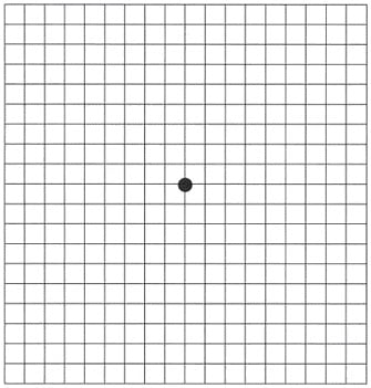
- Application Method of Amsler Grid Test
What is Macular Disease?
Macular degeneration (age-related macular degeneration) is the loss of central vision as a result of the progression of receptor cells in the macula region, which is responsible for clear vision, in the retina layer of the eye.
Macular region< /strong> is located behind our eyes and is the area that allows us to see details. Thanks to the function performed by this region, we can read a book, thread the thread through the eye of a needle, and do many other tasks that require detailed vision.
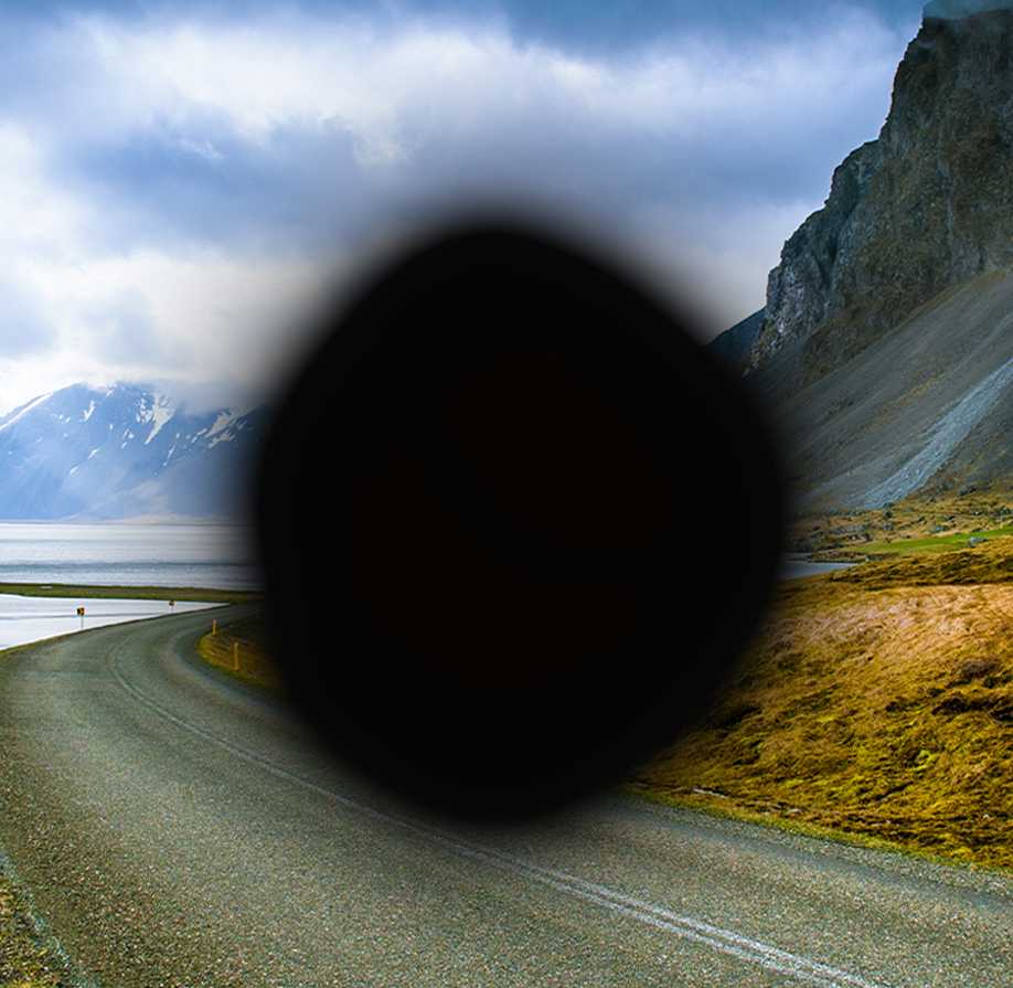
In people with macular degeneration, central vision loss begins as nerve cells die in the center of the visual field. This vision loss begins in the center and expands to the edges over time. Patients see a dark spot in the center and a ring around the edges. As a result of all these, the quality of life of patients is seriously affected.
How is Macular Disease Treated?
Macular macular degeneration for dry type is mostly done by administering special vitamin supplements and high doses of Omega-3, and the entire treatment is carried out with oral medications. Wet type macular degeneration is mostly done by administering anti-VEGF drugs into the eye. It is done by injection. The aim here is to ensure that the disease responds to treatment and to prevent blindness. Many repetitions of these injections may be required.
The treatment of age type of macular degeneration is carried out with drug therapy, intraocular injection therapy and macrovision surgery. Before the surgery. With the simulation, the possible increase in the patient's vision after the surgery can be revealed.
Early diagnosis is important in stopping vision loss and even improving it in certain conditions. After the surgery, the patient's vision can be improved by wearing near and far glasses. Vision loss can be prevented to a certain extent.
MACRO-vision Operation in Yellow Spot Treatment
The surgical method used for the treatment of macular degeneration is macrovision surgery. The central vision of the retina due to various diseases affecting the visual center and indirectly It is an operation that allows patients whose vision is impaired at far and near distances to see better at far and near distances by enlarging the image falling on the retina, without treating their existing diseases. The eye is used like a telescope with the distance and near glasses used after the surgery.
DIAGRAM
 < /p>
< /p>
How Is It Diagnosed?
Macular degeneration can cause various eye diseases and systemic disorders that cause vision loss, especially in older ages. For this reason, the priority for the diagnosis of macular degeneration begins with a detailed listening to the patient's complaints and the medical history of both himself and his family.
The diagnosis of whether the person has the disease is made through an in-depth eye examination by an ophthalmologist. can be revealed. The patient can undergo a detailed retinal examination using a biomicroscope and various lenses and tomography devices. The extent of the visual field can be determined by using different devices.
Thus, deteriorations in the visual field are diagnosed. Depending on the type of deterioration, the presence of macular degeneration can be questioned.
A drop that helps dilate the pupil can be used to make this examination more comfortable. If deemed necessary, the physician may also use a method such as fluorescein angiography, which helps examine the vascular structures in the retina.
What are the precautions that can be taken?
Regular physician check-ups are important to determine early diagnosis of macular degeneration.
Ensure that other health problems are kept under control. For example, if you have a cardiovascular disease, ensure regular medication use without interrupting your doctor's check-ups.
Do not smoke, and if you do, quit. Smoking seriously increases the risk factors for macular degeneration.
Exercise regularly, protect your health and stay at your ideal weight.
Eat a diet rich in fruits and vegetables. These foods contain antioxidants that reduce macular degeneration risk factors.
Consume fish at regular intervals. Omega-3 acids found in fish help reduce the risk of macular degeneration. Walnuts and many other nuts are foods rich in omega-3.
CLICK TO MAKE AN APPOINTMENT WITH OUR EYE DOCTORS
When Should You See a Doctor?
It would be beneficial to see a physician in the following cases:
- You notice darkening in your central vision.
- If you have difficulty seeing colors and fine details, or if you experience problems with straight lines appearing bent.< /li>
These changes may be the first symptoms of macular degeneration, especially if you are over 60 years old. In addition, it would be beneficial for those with a family history of macular degeneration to be more sensitive about certain symptoms.
What are the Symptoms of Macular Disease?
In age-related macular degeneration, cells in the macula, the visual point of the eye, are gradually lost. Therefore, the disease does not suddenly cause very noticeable symptoms. The disease may not be noticed until vision loss occurs. Different symptoms occur in different people. In some cases, symptoms may occur in only one eye, but there is no problem in the other eye.
Macular degeneration becomes easier to diagnose only if both eyes are affected. The initial symptoms are that colors appear faded, text appears blurry, and straight lines begin to appear oblique. As macular degeneration progresses, a dark area forms in the middle of the viewed spot, and problems such as blurring and oblique vision of straight lines increase.
In addition, more light is needed for close reading and fine-detailed work. There is difficulty in adapting to dimly lit environments. As the disease progresses, these problems make it impossible for people to carry out their daily activities alone after a while.
What are the causes of the disease?
The retina layer has its own vascular layer. This layer is still fed by another richer vascular layer underneath it.
There is a thin membrane layer between these two layers that acts as a filter. In the macula region, harmful substances formed by light and other factors are cleared in this membrane layer. The cleaned harmful substances are transmitted to the rich layer below and are eliminated from this region with the help of the circulatory system.
Harmful substances that cannot be eliminated from this region for a reason that is not fully known to date, accumulate in this region and initiate some negative events for the visual system. Thus, macular degeneration begins.
What are the Risk Factors?
Risk factors for macular degeneration are as follows:
- Women,
- People over 65 years of age,
- People with colored eyes,
- Overweight,
- Smoking,
- Those with cardiovascular problems,
are in a higher risk group.
What are the Types of Macular Degeneration?
Age-related macular degeneration has wet and dry types in terms of development type.
Dry Type Macular Degeneration: It is the most common type of the disease. This type is observed in 85% of patients. Small yellow substances called drusen accumulate in the macula region of the retina and damage the macula, thus causing vision loss.
Regular ophthalmologist check-ups are important for the treatment of dry type macular degeneration. Antioxidant-based drug treatment consisting of vitamins and minerals stands out for macular degeneration.
Wet-Type Macular Degeneration: Wet-type macular degeneration occurs in 10-15 percent of patients. species are seen. It occurs as a result of the expansion of abnormal blood vessels under the macula. The macular structure is damaged due to bleeding and extravasation of fluid. This condition may cause vision loss.
Vision loss in this type of the disease can be restored to a certain extent with macrovision treatment for suitable patients.
Apart from these two types, Stargardt< Juvenile type macular degeneration, also known as /strong>, is a type of macular degeneration seen in children and teenagers.
Application Method of Amsler Grid Test
The definitive diagnosis of macular degeneration is made by an ophthalmologist. However, it is a test that may be useful for detecting some symptoms. You can apply the Amsler grid test yourself at home.
- Wear glasses or contact lenses that you normally use for reading.
- Apply the diagram above in a well-lit room, approximately 30 – 40 meters away from your face. Look at it from a distance of cm.
- Cover one eye with your hand and focus on the point in the middle with your open eye. Make sure that you can see all 4 corners of the large square in the diagram.
- Test your other eye in the same way.
- If you see ripples, breaks, blurriness in the lines, or if you cannot see the corners, these are yellow eyes. There may be symptoms of spot disease.
- In this case, it is necessary to consult an ophthalmologist who specializes in retinal diseases as soon as possible.

Published Date: 04/03/2024
Updated Date: 04/03/2024