Cornea and Diseases
Cornea is a transparent and dome-shaped tissue located in the outermost layer of the eye, in front of the iris and pupil. It protects the structures inside the eye and contributes to the healthy reflection of the image on the retina by performing the first refraction of light coming into the eye from outside. It is the layer with the highest refractivity in our eyes. For this reason, even the smallest problem in the cornea significantly affects vision.
SUBJECT TITLES
- What is the Structure of the Cornea?
- Conjunctivitis and its Treatment
- Corneal Ulcer and Treatment
- Corneal Dystrophies and Treatment
- Cornea Transplantation (Keratoplasty)
- Keratoconus and its Treatment
- Keratitis and its Treatment
- Microcornea and its Treatment
- Corneal Diseases and How Are They Treated?
- How Are Corneal Diseases Diagnosed?
- What are the Cornea Functions?
- Pterygium and its Treatment
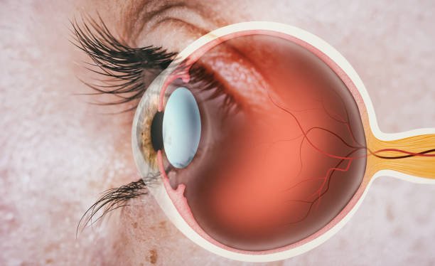
What is the Structure of the Cornea?
The horizontal diameter of a healthy cornea is approximately 12 millimeters. When viewed from the front, its vertical diameter is 11 millimeters. Its average thickness is 550 microns. Its parts are as follows:
- Corneal Epithelium: It is the outermost layer. It is around 50 microns and consists of 5 layers of cells. Epithelial cell production occurs in the surface tear layer. These cells continually move upwards, replacing older cells. This change occurs continuously, on average, within 1 week.
- Bowman's Layer: It is formed by the compression of collagen fibers. It is the transition layer between the epithelial and stroma layers. It has a very thin structure.
- Corneal Stroma:It constitutes the majority of the entire corneal thickness and is the middle layer. It consists of collagen fibers. When damaged, it usually results in loss of transparency and changes in curvature, resulting in serious vision loss.
- Descement Membrane: It separates the stroma layer from the endothelial layer. It has a very thin structure. It gradually thickens over time. As a result of its damage, the corneal endothelium is damaged.
- Corneal Endothelium: It is the innermost layer. It is only a single cell layer thick and is around 5 microns thick. It has a semi-permeable structure. Most of the endothelial cells in this layer have a hexagonal structure and the arrangement of these cells is also called endothelial mosaic.
Conjunctivitis and its Treatment
Conjunctivitis is one of the diseases that can directly affect this area, even though it does not appear directly on the corneal surface. Conjunctivitis is also commonly known as red eye disease. External factors play an important role in the emergence of the disease. Conjunctiva is a network layer with very thin vessels in its internal structure, and its main function is to keep the upper surface of the eye moist.
Conjunctivitis is the inflammation of the conjunctiva layer, which is the outermost white part of the eyeball, due to reasons such as bacteria, allergies and viruses. Sometimes it can be seen in one eye, and sometimes it can be seen in both eyes. The disease is contagious in some cases. Apart from its occurrence due to allergies, it may also occur due to non-compliance with hygiene rules.
Conjunctivitis treatment varies depending on the type of disease. Methods such as eye drops and hot compresses are often used. Especially in viral conjunctivitis, the patient is expected to pay attention to hygiene rules.
Corneal Ulcer and Treatment
Corneal ulcer occurs with symptoms of eye redness, mild eye discharge and vision loss. An ulcer usually results from an abscess-type surface infection. It can also occur as a result of eye injuries and various traumas.
Other diseases that can affect the cornea are conjunctivitis and pterygium.
Corneal Dystrophies and Treatment
It is a disease of genetic origin that can occur in the corneal layer. It can be seen in both eyes. Depending on the type of genetic mutation that occurs and its location in the cornea, some accumulations that may cause clouding may be observed. Depending on the location and density of these deposits, blurred vision and vision loss occur.
If the deposits are in the frontal area, symptoms such as stinging, burning and pain in the eyes may occur. Depending on the type of dystrophin that occurs, full corneal transplantation or partial transplantation may occur.
Cornea Transplantation (Keratoplasty)
Cornea transplantIt involves removing the corneal layer that has lost its transparency and formal integrity and transplanting the healthy tissue with a surgical procedure. The transplantation process includes the transplantation of the inner or upper part and the full cornea transplantation. After a successful procedure, vision is greatly improved.
The situations in which transplantation is applied are as follows:
Extensive deterioration in vision and deformities that occur when keratoconus and various corneal diseases cannot be corrected with medication and surgical intervention. In cases where it occurs,
Loss of transparency of the cornea as a result of edema in this layer and corneal edema that causes permanent damage as a result of keratitis,
Deformity that occurs with swelling in the cornea as a result of bullous keratopathy,
p>
Due to traumatic and foreign object-related injuries on the surface,
the need for transportation may arise.
Keratoconus and its Treatment
Keratoconus is a corneal disease in which the structure of the cornea deteriorates, becomes thinner and bulges towards the front, with astigmatism and myopia tending to progress. It is progressive, and in cases where it occurs in both eyes, it may progress more in one eye than the other. It usually starts in adolescence and progresses with the age of 20.
The disease shows mild blurring of vision in the initial stage. Fluctuations and curvatures in the appearance of straight lines and frequent changes in eye numbers are the most common symptoms.
Keratoconus treatment varies depending on the level of the disease. It is a multi-stage treatment. In the early stages of the disease, vision disorders can be corrected with the use of glasses and contact lenses. In advanced stages of the disease, treatment can be applied with hard contact lenses and scleral lenses. However, all these methods cannot stop the progression of the disease permanently. Permanent progression of the disease can only be stopped by corneal cross-linking treatment. Cross-linking treatment and hybrid treatment are performed on suitable patients. In hybrid treatment, corneal crosslinking treatment and No Touch Laser are applied together. In very advanced cases where all these treatments do not work, corneal transplantation may be possible.

Keratitis and its Treatment
Keratitis is the inflammation of the cornea, the outermost surface of the eye. Corneal tissue may become cloudy due to keratitis, and in later cases, perforations may occur in this tissue. Inflammation can occur with or without infection. If the cause of the disease is not due to infection, it may occur due to long-term use of contact lenses and foreign objects coming into contact with the eye. If it is due to infection, it may occur due to bacteria, viruses, etc.
It shows symptoms in the form of pain and stinging in the eye, redness and blurred vision, as well as white spots on the corneal surface. Treatment of keratitis due to infection is carried out according to the intensity and type of infection. Keratitis caused by bacteria is treated with eye drops. Antibiotics can be used in keratitis caused by an advanced infection.
Microcornea and its Treatment
Microcornea is defined as a cornea with a horizontal diameter of less than 10 mm. As a result of this problem, it reduces the refraction of light coming from outside. Treatment can be done by surgical methods.
Corneal Diseases and How Are They Treated?
Cornea treatment varies depending on the type and level of diseases occurring in this region.
The determining factor for cornea treatment is the type and level of the disease. Treatment begins after the corneal disease is diagnosed through various tests. Early diagnosis for corneal treatment is important for the success of the treatment, as in other eye diseases.

How Are Corneal Diseases Diagnosed?
Corneal diseases are diagnosed through various tests performed by the ophthalmologist in addition to the general eye examination. Each examination is aimed at evaluating different segments of the structure of this layer. As a result of various examinations, diseases existing in the cornea can be revealed. As with all eye health-related diseases, early diagnosis is important for the success of treatment.
What are the Cornea Functions?
Cornea is located at the front of the eye and is the first area where light from outside enters the eye. The cornea is the outermost refractive component of the eye and the other component is the natural eye lens. The light coming from outside first passes through this region and then through the eye lens and focuses on the retina. When this process functions in a healthy way, visual function occurs. The transparency and shape of the cornea must be perfect in order to avoid any loss of vision during the process, that is, in order to refraction the light coming from outside in the most ideal way. Because this region provides more than half of the focusing power of the eye. The refractive power of the cornea is stable, not variable, like the natural intraocular lens.
The other function of the cornea layer, apart from providing refraction of light coming from outside, is to protect the inner part of the eye against dust, microbes and various foreign substances coming from outside. It performs this function together with the eyelids, tears and sclera tissue. It contributes to the protection of the retina by acting as a filter against the harmful effects of sunlight.
Pterygium and its Treatment
It is also known as pterygium eyelid. Pterygium occurs in a thin-vascular structure, but as it progresses, it thickens and attaches to the corneal layer, causing traction there, causing astigmatism and blurred vision. As it progresses to the pupil, it can completely block the view of outside light, causing permanent vision loss. Surgery is the only permanent method to correct the pterygium problem. It may be necessary to take precautions to prevent the disease from recurring in the future.

Published Date: 04/03/2024
Updated Date: 18/03/2024