What is the Eye and What is the Eye Anatomy?
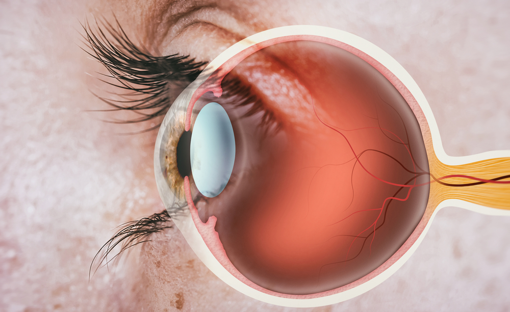
Eye anatomy is of great importance in terms of protecting eye health and diagnosing and treating eye-related problems. The eye is one of the most important sensory organs of the human body and has a complex structure that enables the visual process. Eye anatomy consists of 3 parts: outer layer (sclera and cornea), middle layer (choroid, iris, ciliary and cornea) and inner layer (retina). The ability to see is gained thanks to the interaction of these complex structures. Components such as cornea, iris, retina and optic nerve play critical roles in the functioning of the eye anatomy. Understanding eye anatomy is of great importance to protect eye health and detect visual disorders. The complexity of eye anatomy is carefully examined by ophthalmologists for healthy vision and the detection of diseases. Regular examinations are important for protecting eye health and early diagnosis of visual disorders.
SUBJECT TITLES
- What is the Eye?
- Structure of the Eye: Main Parts and Their Functions
- Cornea: Transparent Window of the Eye
- Iris and Pupil: The Control Center of Light
- The Lens: The Key to Image Creation
- Retina: Where Light Turns into Image
- Optic Nerve: Image Transmission to the Brain
- Intraocular Pressure and Glaucoma: Aqueous and Vitreous Humor
- Source
What is the Eye?
The eye functions as the visual organ of humans and other living things. The eye detects environmental stimuli and initiates the vision process through signals sent to the brain.
Structure of the Eye: Main Parts and Their Functions
The eye is the body structure used by living organisms to perceive the light around them, create images and transmit them to the brain. Eye anatomy has a complex structure. Thanks to the optical system and neural connections, it provides the vision process by transmitting the perceived light and visual information to the brain. The structures in the eye anatomy are as follows:
- Cornea:The transparent structure that forms the outer layer of the eye anatomy.
- Sclera: The hard and durable tissue layer that forms the white part.
- Conjunctiva:The structure located between the sclera and the eyelid and forming the inner part of the eyelid.
- Iris. :It is the colored part of the eye anatomy that includes the muscles that determine eye color and regulate the pupil.
- Lens:It is a transparent, flexible structure and allows focusing.
- Retina:It is the innermost layer of the eye anatomy and contains photoreceptor cells to convert light into electrical signals and transmit it to the brain.
- Optic Nerve:Nerve fibers that transmit signals from the retina to the brain.
- Eye Muscles:These are the muscles that control movement in the eye anatomy.
- Eyelids:With their eyelashes, they protect against foreign objects. It has a protective effect.
- Tear Glands:Protects and moisturizes the eyes. It has a protective effect against foreign objects.
Cornea: Transparent Window of the Eye
Cornea is a transparent and dome-shaped structure that forms the outer layer of the eye anatomy. It lies above other structures such as the iris and lens and covers the front part of the eye. The cornea acts as a protective layer from environmental influences and prevents damage to the internal parts. The cornea has a complex structure consisting of many layers. It consists of epithelium, Bowman, stroma, Descemet and endothelial layers from outside to inside. A smooth and healthy cornea helps to create clear and sharp images by refracting light correctly. This is the first step in the vision process and helps create images that are reflected in reverse on the retina.
The cornea has a transparent structure. Because there are almost no blood vessels between the cells in the cornea. The absence of blood vessels preserves the transparency of the cornea by preventing blood leakage into the eye. At the same time, the regular cell structure and arranged collagen fibers of the cornea also allow light to pass freely. In addition, the cornea is one of the most sensitive areas in the eye anatomy. It is the first structure of the eye anatomy to be exposed to irritating foreign objects and triggers reactions such as the blink reflex to protect the surface.
Iris and Pupil: The Control Center of Light
Iris is the name of the ring-shaped structure that forms the colored part of the eye. It determines eye color and regulates its size by surrounding the pupil. Different colors such as blue, green, brown are determined depending on the levels of melanin in the iris. The pupil is the dark round opening located in the middle of the iris. Its size changes depending on the amount of light. The iris muscles surrounding the pupil control this process of growth and contraction. The task of the pupil is to regulate the amount of light entering and to create a clear image by controlling the amount of light falling on the retina.
The iris and pupil work together to provide light control. When there is little light, the pupil expands and tries to let in more light. However, under very bright light, the baby shrinks due to eye anatomy and prevents the harmful effects of light by preventing excess light from entering the eye.
The Lens: The Key to Image Creation
The lens is a structure located in the back part of the eye anatomy, between the iris and the retina. It has a transparent and elastic structure, approximately 9-10 mm in diameter and 4 mm in thickness. The main function of the lens is to create a sharp image on the retina by refracting and focusing incoming rays inwards. The lens corrects the image reflected upside down on the retina in the correct direction. This process ensures that the image perceived by the photoreceptor cells on the retina is clear and sharp. The power provided through the zonular fibers helps change the shape of the lens. When the eye looks at distant objects, the lens flattens, refracting light less and creating a clear image. However, when the eye looks at close objects, the lens thickens and refracts the light more, providing a clear image. Health problems that may occur in the lens are:
Retina: Where Light Turns into Image
Retina is located in the back part of the eye anatomy. It is one of the most important components of the vision process, which begins with light falling on the eye. The light detected by the fundus is perceived as an image on the retina. The light focused by the lens falls on the photoreceptor cells in the retina. These photoreceptors convert light into electrical signals and transmit them to the brain. These signals, sent to the brain through the optic nerve originating from the part called the blind spot, help perceive and interpret the image. In the inner part of the retina, there is a region containing two photoreceptor cells, cones and rods, where visual information is perceived. The rods are effective in low light levels and night vision. They cannot perceive color and have lower resolution. Cones, on the other hand, are effective at higher light levels and color vision. They perceive details by providing good resolution. There are areas on the retina where cone cells called macula are concentrated and where vision is clearest, called fovea. In eye anatomy, the layer located just below the retina and supporting photoreceptor cells is the retinal pigment epithelium.
Optic Nerve: Image Transmission to the Brain
The optic nerve is a bundle of nerves that starts from the back of the eye anatomy and extends to the brain. Nerve cells located at the back of the retina form the beginning of the optic nerve. These nerve cells collect information from photoreceptor cells (rods and cones) that convert light into electrical signals and transmit the information along the optic nerve to be transmitted to the brain. The optic nerve follows a path that starts from the back of the eye anatomy and extends to the visual centers in the brain. This path eventually crosses at a point behind the eye. This crossover, called the optic chiasm, mixes the image information from the right and left eyes and enables the information in the visual fields of both eyes to come together. The resulting neural pathway then proceeds to different vision centers in the brain and transmits visual signals to the regions necessary for processing and interpreting visual information.
Intraocular Pressure and Glaucoma: Aqueous and Vitreous Humor
There are different fluids between the front and back parts of the eye anatomy. There are fluids called "aqueous humor" produced by the choroidal ciliary in the front part and "vitreous humor" in the back part. Aqueous humor is a transparent fluid located in the front part of the eye anatomy and is located in the section called the anterior chamber. It plays an important role in preserving the transparency and shape of the front section. Aqueous humor is constantly produced and kept in balance by being absorbed through the tear ducts. This fluid contributes to the regulation of internal eye pressure and protects the mechanical structure. Vitreous humor is a gel-like fluid located at the back of the eye anatomy and is located in the section called the posterior chamber. Vitreous humor supports the shape of the retina at the back of the eye, helping to perceive visual information clearly. It also protects the mechanical structure of the eye and ensures proper placement of the retina. Intraocular pressure is kept under control thanks to the balance and circulation of these fluids. However, in some cases, the pressure in the eye anatomy increases and can cause a serious disorder known as glaucoma.
Glaucoma, pressure It is a disease that occurs as a result of elevation and damage to nerve fibers. High pressure occurs especially in situations that prevent the drainage of aqueous humor. This situation causes pressure on the optic nerve over time.
Regular examinations are of great importance to ensure the healthy functioning of the eye anatomy and to detect problems related to the eye anatomy at an early stage.
Source
https://www.britannica.com/science/human-eye
https://www.aao.org/eye-health/anatomy/parts-of-eye https://www.ncbi.nlm.nih.gov/books/NBK11120/ https://www. ncbi.nlm.nih.gov/books/NBK11120/
Published Date: 31/07/2023
Updated Date: 04/03/2024